
Diagnostics
Dental Diagnostics: How Do Digital X-Rays Work, and Why Are They Done?
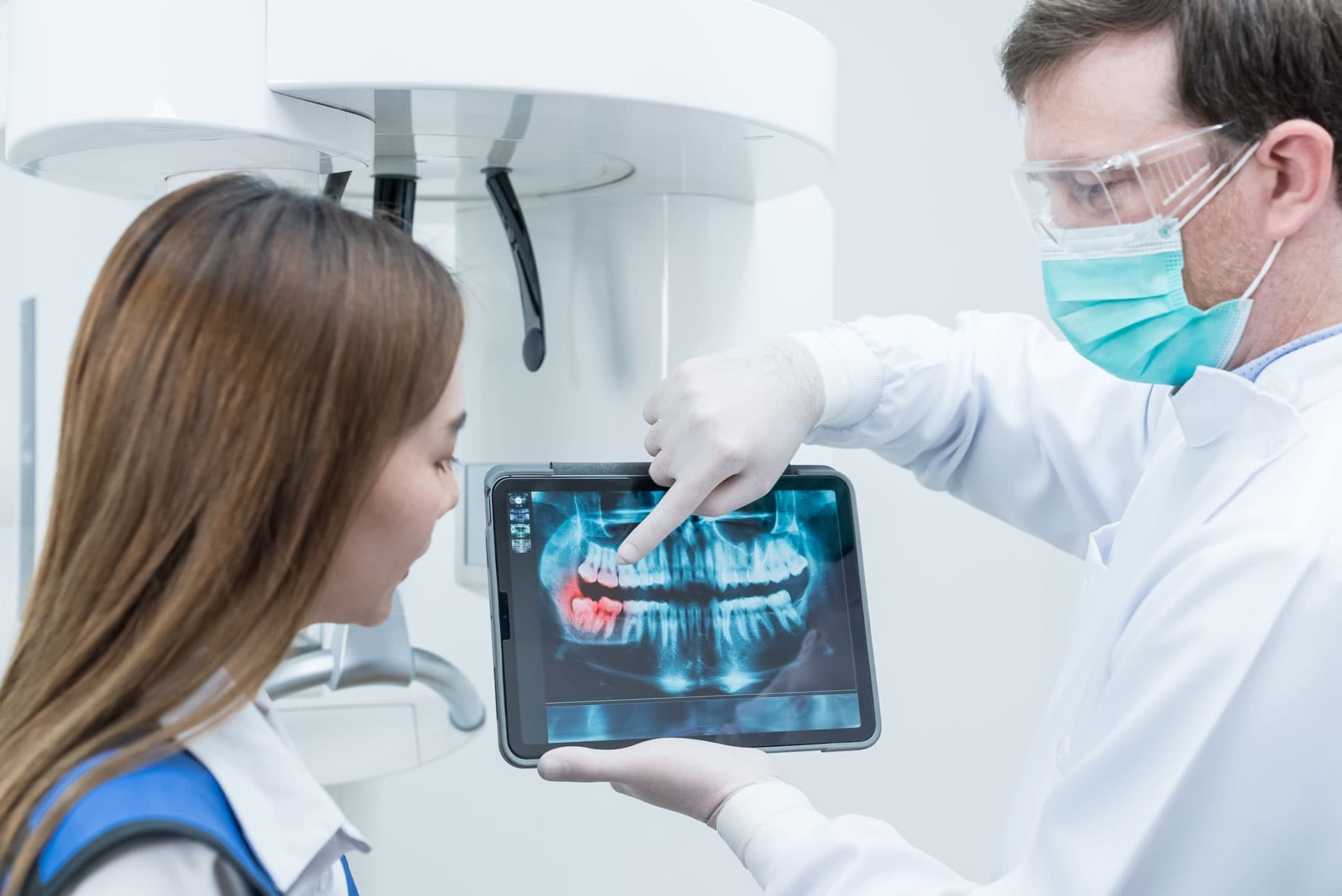
X-rays help dentists evaluate areas that are difficult to see during an examination, including between teeth and around their roots. Digital images support diagnosis, but do not replace an examination or a discussion of your symptoms.
Digital X-rays in dental diagnostics
Digital radiography uses an electronic sensor or a scanned imaging plate instead of photographic film. The image can be viewed on a computer and stored with your dental records.
The benefits of digital X-rays
Moving from film to digital changed dental diagnostics in several meaningful ways.
Images that support diagnosis
Image quality depends on the equipment, positioning, and technique. Digital imaging is not a guarantee that every problem will be visible.
A closer look
Digital images can be enlarged on screen to help your dentist review an area and explain the findings to you.
Immediate viewing
There is no waiting for photographic film to develop. Once the digital image is available, your dentist can review it with you. Further tests or a separate treatment appointment may still be needed.
Spotting the unseen
Radiographs provide information about teeth and surrounding bone that complements the clinical examination. Findings may lead to further evaluation rather than a diagnosis from the image alone.
Planning the next step
Ask how the findings affect your options, what needs attention now, and what can be monitored. Discuss the proposed treatment and its cost before deciding on the next step.
Understanding radiation exposure
Digital imaging still uses ionizing radiation. Exposure varies by examination and technique; a single percentage reduction does not describe every comparison with film.
Electronic storage and easy access
Digital images can be stored electronically. Ask the office how to request copies or arrange for records to be transferred to another dental provider.
A more sustainable choice
Digital X-rays do not require the chemicals used to develop film, so they are gentler on the environment. At Southern Smiles, we care about reducing our footprint, and this technology helps us do that.
The technology behind digital X-rays
Digital X-ray technology, also called digital radiography, has reshaped dental imaging. Here is what makes it work.
The digital sensor
At the heart of the system is a small digital sensor. It is placed in your mouth to capture the images. Rather than relying on film that must be developed, the sensor captures X-ray energy and converts it directly into electronic signals.
The software
Those signals travel to a computer, where dedicated software interprets them and builds a visual image. Because the process is computerized, your dental team can use image enhancement tools to highlight anything that needs a closer look.
The monitor
The finished images appear on a monitor right away. This saves time and helps with communication, since your dentist can review the images with you and talk through any findings on the spot.
Keeping X-rays safe
Digital X-rays involve radiation exposure. Ask why an image is recommended, how it will inform your care, and whether previous images are available. Tell your dental team about your medical history and any pregnancy or other concerns before imaging.
Types of digital dental X-rays and their uses
Digital dental X-rays come in different types, each designed to capture a specific view of your teeth and mouth. The type your dentist chooses depends on what they need to see or monitor.
Bitewing X-rays
Bitewing X-rays are named for the small plastic bite tabs you hold between your teeth while the image is captured. They show the upper and lower back teeth in a single view. They help your dentist find cavities between teeth, spot changes in bone density from gum disease, and check the fit of a crown or other restoration.
Periapical X-rays
Periapical X-rays focus on one or two whole teeth, from the root to the crown. They are useful when your dentist needs a detailed view of a specific tooth. They can reveal problems in the root structure and surrounding bone, help diagnose an abscess, or track the progress of a treatment.
Panoramic X-rays
Panoramic X-rays give a broad view of the entire mouth in one image. They capture the teeth, upper and lower jawbones, sinuses, and temporomandibular joints (TMJ). Because of that wide view, they are often used to plan treatments such as implants, braces, dentures, or extractions. They also help identify fractures, cysts, tumors, bone irregularities, and infections in the jaw.
Making the most of your digital X-ray
Open, proactive communication with your dentist gets you the most out of any X-ray. Ask questions, understand the process, and know what the results may mean. Following the instructions you are given, and reporting any changes in your dental health, helps too.
Why choose Southern Smiles for your dental care
At Southern Smiles in South Phoenix, ask Dr. Monzer Shakally what information an image would add to your examination and treatment plan. If your visit concerns a painful tooth, explore our emergency dental care and root canal information. You can also review our treatment plans and costs FAQs before your visit.
Do I need X-rays at every visit?
Not automatically. The decision depends on your examination, health history, risk factors, and previous images. Ask whether existing records can answer the clinical question before repeating an examination.
For background, see the American Dental Association's guidance on dental radiographs and its 2026 patient-selection recommendations.
Ready for a healthier smile?
We provide a thorough exam as part of every wellness visit. Book an appointment and let us take care of you.